Big news! We're excited to announce our brand new product, LabTab. Learn more

Works citing SciDraw

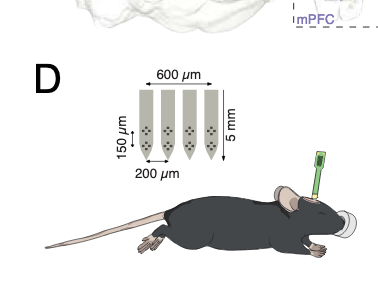
Network asynchrony underlying increased broadband gamma power

N. Guyon, L. R. Zacharias, E. Fermino de Oliveira, H. Kim, J. Pereira Leite, C Lopes-Aguiar, M Carlén

Synchronous activity of cortical inhibitory interneurons expressing parvalbumin (PV) underlies the expression of cortical gamma rhythms. Paradoxically, deficient PV inhibition is associated with increased broadband gamma power. Increased baseline broadband gamma is also a prominent characteristic in schizophrenia, and a hallmark of network alterations induced by N-methyl-D-aspartate receptor (NMDAR) antagonists such as ketamine. It has been questioned if enhanced broadband gamma power is a true rhythm, and if rhythmic PV inhibition is involved or not. It has been suggested that asynchronous and increased firing activities underlie broadband power increases spanning the gamma band. Using mice lacking NMDAR activity specifically in PV neurons to model deficient PV inhibition, we here show that local LFP (local field potential) oscillations and neuronal activity with decreased synchronicity generate increases in prefrontal broadband gamma power. Specifically, reduced spike time precision of both local PV interneurons and wide-spiking (WS) excitatory neurons contribute to increased firing rates, and spectral leakage of spiking activity (spike “contamination”) affecting the broadband gamma band. Desynchronization was evident at multiple time scales, with reduced spike-LFP entrainment, reduced cross-frequency coupling, and fragmentation of brain states. While local application of S(+)-ketamine in wildtype mice triggered network desynchronization and increases in broadband gamma power, our investigations suggest that disparate mechanisms underlie increased power of broadband gamma caused by genetic alteration of PV interneurons, and ketamine-induced power increases in broadband gamma. Our studies, thus, confirm that broadband gamma increases can arise from asynchronous activities, and demonstrate that long-term deficiency of PV inhibition can be a contributor.
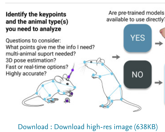
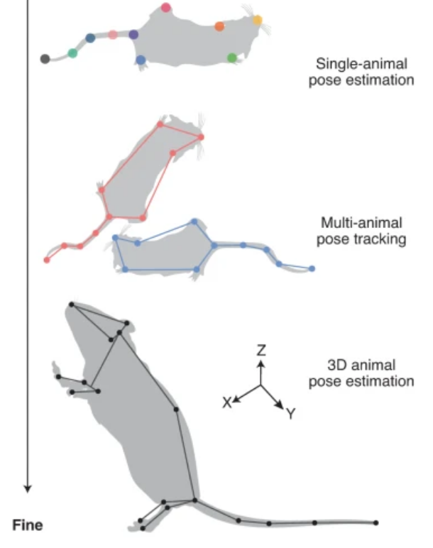
A Primer on Motion Capture with Deep Learning: Principles, Pitfalls, and Perspectives

A. Mathis, S. Schneider, J. Lauer, M. W. Mathis

Extracting behavioral measurements non-invasively from video is stymied by the fact that it is a hard computational problem. Recent advances in deep learning have tremendously advanced our ability to predict posture directly from videos, which has quickly impacted neuroscience and biology more broadly. In this primer, we review the budding field of motion capture with deep learning. In particular, we will discuss the principles of those novel algorithms, highlight their potential as well as pitfalls for experimentalists, and provide a glimpse into the future.

Control of Orienting Movements and Locomotion by Projection-Defined Subsets of Brainstem V2a Neurons

G. Usseglio, E. Gatier, A. H, C. Hérent, J. Bouvier

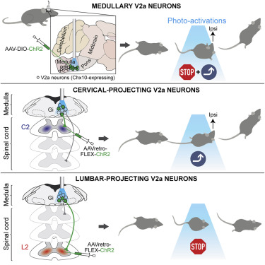
Spatial orientation requires the execution of lateralized movements and a change in the animal’s heading in response to multiple sensory modalities. While much research has focused on the circuits for sensory integration, chiefly to the midbrain superior colliculus (SC), the downstream cells and circuits that engage adequate motor actions have remained elusive. Furthermore, the mechanisms supporting trajectory changes are still speculative. Here, using transneuronal viral tracings in mice, we show that brainstem V2a neurons, a genetically defined subtype of glutamatergic neurons of the reticular formation, receive putative synaptic inputs from the contralateral SC. This makes them a candidate relay of lateralized orienting commands. We next show that unilateral optogenetic activations of brainstem V2a neurons in vivo evoked ipsilateral orienting-like responses of the head and the nose tip on stationary mice. When animals are walking, similar stimulations impose a transient locomotor arrest followed by a change of trajectory. Third, we reveal that these distinct motor actions are controlled by dedicated V2a subsets each projecting to a specific spinal cord segment, with at least (1) a lumbar-projecting subset whose unilateral activation specifically controls locomotor speed but neither impacts trajectory nor evokes orienting movements, and (2) a cervical-projecting subset dedicated to head orientation, but not to locomotor speed. Activating the latter subset suffices to steer the animals’ directional heading, placing the head orientation as the prime driver of locomotor trajectory. V2a neurons and their modular organization may therefore underlie the orchestration of multiple motor actions during multi-faceted orienting behaviors.

Quantifying behavior to understand the brain

T. D. Pereira, J. W. Shaevitz & M. Murthy

Over the past years, numerous methods have emerged to automate the quantification of animal behavior at a resolution not previously imaginable. This has opened up a new field of computational ethology and will, in the near future, make it possible to quantify in near completeness what an animal is doing as it navigates its environment. The importance of improving the techniques with which we characterize behavior is reflected in the emerging recognition that understanding behavior is an essential (or even prerequisite) step to pursuing neuroscience questions. The use of these methods, however, is not limited to studying behavior in the wild or in strictly ethological settings. Modern tools for behavioral quantification can be applied to the full gamut of approaches that have historically been used to link brain to behavior, from psychophysics to cognitive tasks, augmenting those measurements with rich descriptions of how animals navigate those tasks. Here we review recent technical advances in quantifying behavior, particularly in methods for tracking animal motion and characterizing the structure of those dynamics. We discuss open challenges that remain for behavioral quantification and highlight promising future directions, with a strong emphasis on emerging approaches in deep learning, the core technology that has enabled the markedly rapid pace of progress of this field. We then discuss how quantitative descriptions of behavior can be leveraged to connect brain activity with animal movements, with the ultimate goal of resolving the relationship between neural circuits, cognitive processes and behavior.

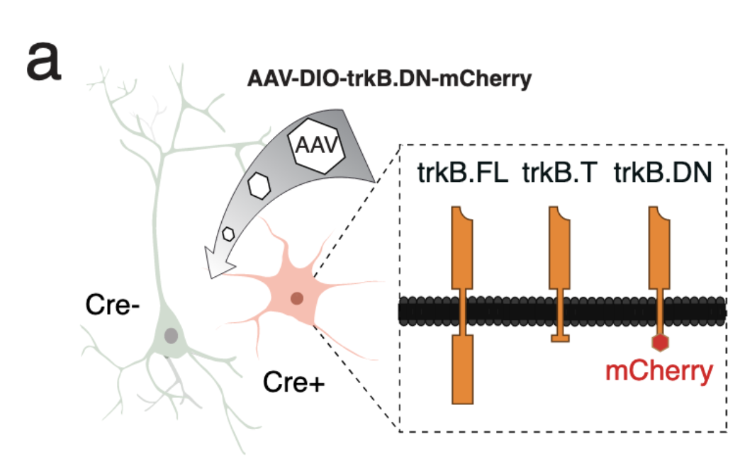
A critical role for trkB signaling in the adult function of parvalbumin interneurons and prefrontal network dynamics

N, Guyon, L. R. Zacharias, J. A. van Lunteren, J. Immenschuh, J. Fuzik, A. Märtin, Y Xuan, M Zilberter, H Kim, K Meletis, C Lopes-Aguiar, MCarlén

Inhibitory interneurons expressing parvalbumin (PV) in the prefrontal cortex (PFC) are central to excitatory/inhibitory (E/I) balance, generation of gamma oscillations, and cognition. Dysfunction of PV interneurons disrupts information processing and cognitive behavior. Tyrosine receptor kinase B (trkB) signaling is known to regulate the differentiation and maturation of cortical PV interneurons during development, but is also suggested to be involved in the activity and network functions of PV interneurons in the adult brain. Using a novel viral strategy for cell-type and region-specific expression of a dominant negative trkB in adult mice, we show that reduced trkB signaling in PV interneurons in the PFC leads to pronounced morphological, physiological, and behavioral changes. Our results provide evidence for a critical role of trkB signaling in the function of PV interneurons in the adult brain, local network activities central to prefrontal circuit dynamics, and cognitive behavior.

A cortico-collicular circuit for accurate orientation to shelter during escape

R. Vale, D. Campagner, P. Iordanidou, O. Arocas, Y. Tan, V. Stempel, S. Keshavarzi, R. S. Petersen, T. W. Margrie, T. Branco

When faced with predatorial threats, escaping towards shelter is an adaptive action that offers long-term protection against the attacker. From crustaceans to mammals, animals rely on knowledge of safe locations in the environment to rapidly execute shelter-directed escape actions1–3. While previous work has identified neural mechanisms of instinctive escape4–9, it is not known how the escape circuit incorporates spatial information to execute rapid and accurate flights to safety. Here we show that mouse retrosplenial cortex (RSP) and superior colliculus (SC) form a monosynaptic circuit that continuously encodes the shelter direction. Inactivation of SC-projecting RSP neurons decreases SC shelter-direction tuning while preserving SC motor function. Moreover, specific inactivation of RSP input onto SC neurons disrupts orientation and subsequent escapes to shelter, but not orientation accuracy to a sensory cue. We conclude that the RSC-SC circuit supports an egocentric representation of shelter direction and is necessary for optimal shelter-directed escapes. This cortical-subcortical interface may be a general blueprint for increasing the sophistication and flexibility of instinctive behaviours.

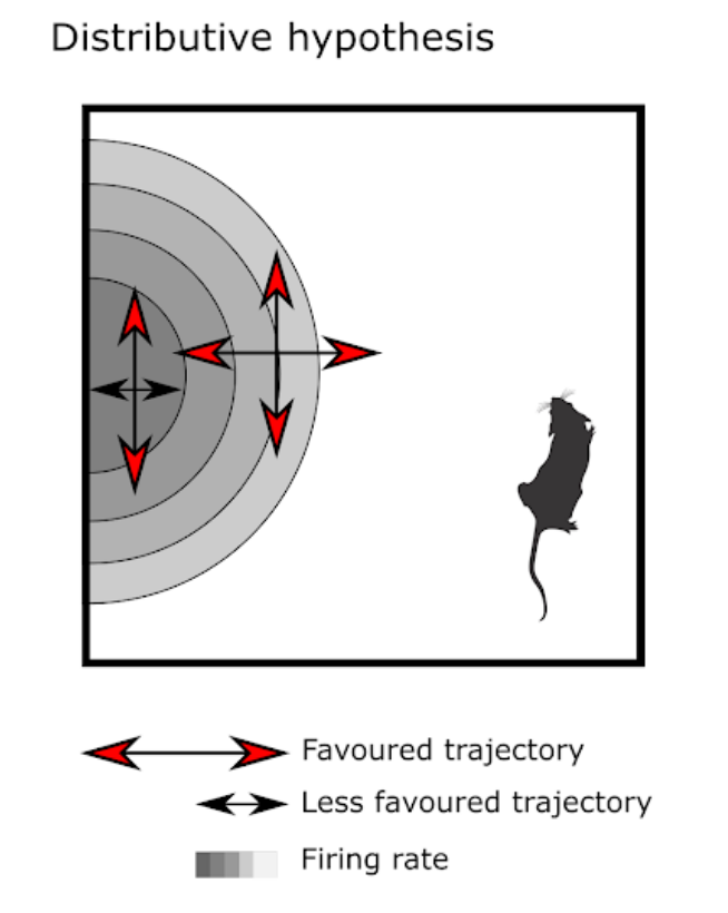
Grid cells encode local head direction

Klara Gerlei, Jessica Passlack, Ian Hawes, Brianna Vandrey, Holly Stevens, Ioannis Papastathopoulos, Matthew F. Nolan

Grid and head direction codes represent cognitive spaces for navigation and memory. Pure grid cells generate grid codes that have been assumed to be independent of head direction, whereas conjunctive cells generate grid representations that are tuned to a single head direction. Here, we demonstrate that pure grid cells also encode head direction, but through distinct mechanisms. We show that individual firing fields of pure grid cells are tuned to multiple head directions, with the preferred sets of directions differing between fields. This local directionality is not predicted by previous continuous attractor or oscillatory interference models of grid firing but is accounted for by models in which pure grid cells integrate inputs from co-aligned conjunctive cells with firing rates that differ between their fields. Local directional signals from grid cells may contribute to downstream computations by decorrelating different points of view from the same location

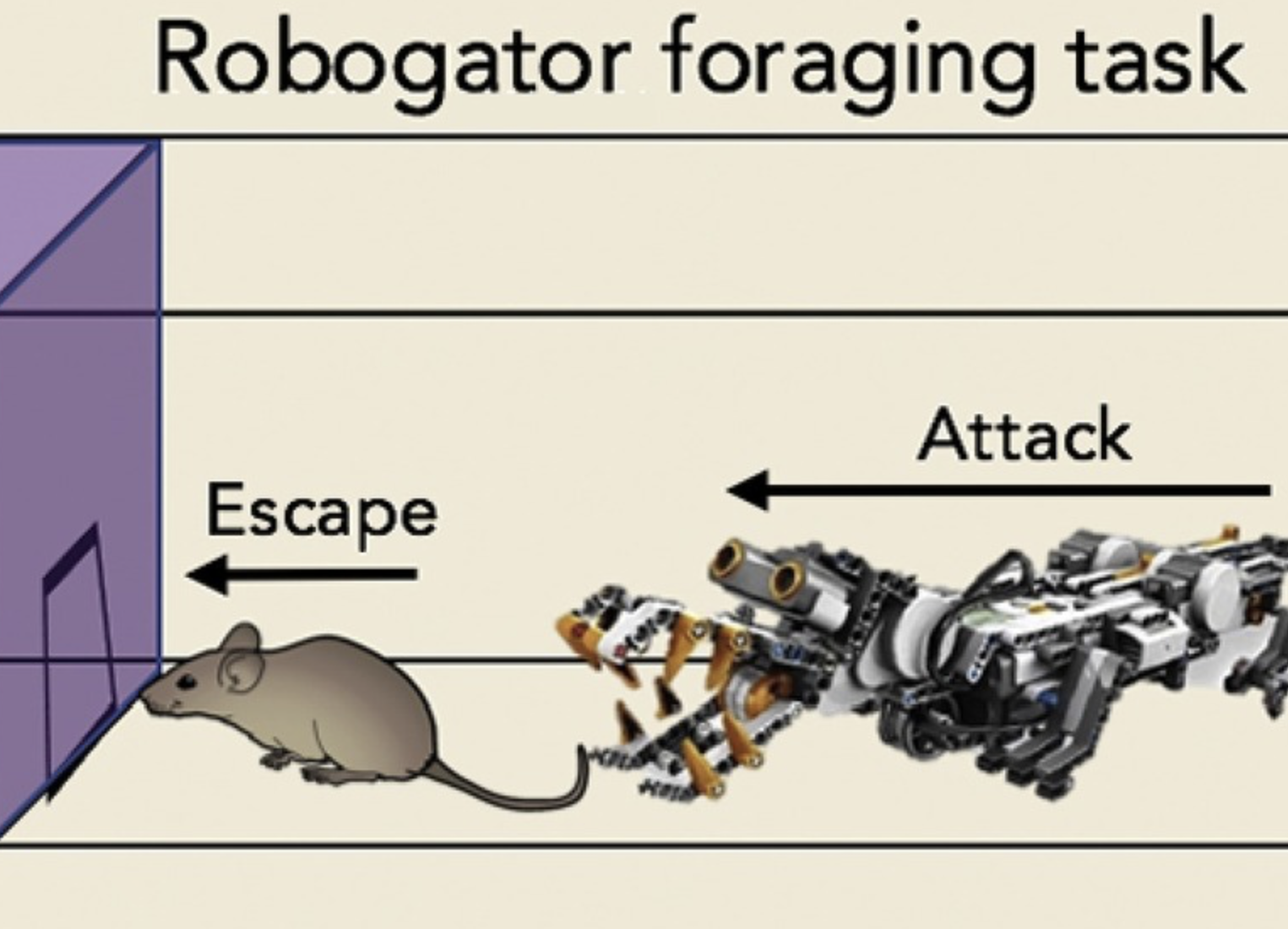
Space, Time, and Fear: Survival Computations along Defensive Circuits

Dean Mobbs, Drew B. Headley, Weilun Ding, Peter Dayan

Naturalistic observations show that decisions to avoid or escape predators occur at different spatiotemporal scales and that they are supported by different computations and neural circuits. At their extremes, proximal threats are addressed by a limited repertoire of reflexive and myopic actions, reflecting reduced decision and state spaces and model-free (MF) architectures. Conversely, distal threats allow increased information processing supported by model-based(MB) operations, including affective prospection, replay, and planning. However, MF and MB computations are often intertwined, and under conditions of safety the foundations for future effective reactive execution can be laid through MB instruction of MF control. Together, these computations are associated with distinct population codes embedded within a distributed defensive circuitry whose goal is to determine and realize the best policy.

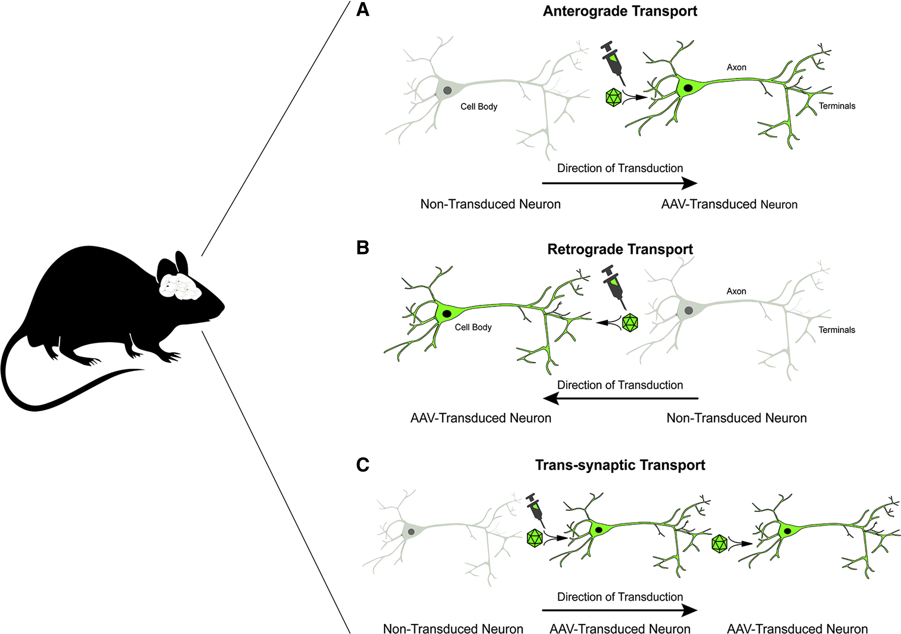
Adeno-Associated Viral Vectors in Neuroscience Research

David L. Haggerty, Gregory G. Grecco, Kaitlin C. Reeves, Brady Atwood

Adeno-associated viral vectors (AAVs) are increasingly useful preclinical tools in neuroscience research studies for interrogating cellular and neurocircuit functions and mapping brain connectivity. Clinically, AAVs are showing increasing promise as viable candidates for treating multiple neurological diseases. Here, we briefly review the utility of AAVs in mapping neurocircuits, manipulating neuronal function and gene expression, and activity labeling in preclinical research studies as well as AAV-based gene therapies for diseases of the nervous system. This review highlights the vast potential that AAVs have for transformative research and therapeutics in the neurosciences.

Hippocampal Remapping as Hidden State Inference

Honi Sanders, Matthew A. Wilson, Samuel J. Gershman

Cells in the hippocampus tuned to spatial location (place cells) typically change their tuning when an animal changes context, a phenomenon known as remapping. A fundamental challenge to understanding remapping is the fact that what counts as a “context change” has never been precisely defined. Furthermore, different remapping phenomena have been classified on the basis of how much the tuning changes after different types and degrees of context change, but the relationship between these variables is not clear. We address these ambiguities by formalizing remapping in terms of hidden state inference. According to this view, remapping does not directly reflect objective, observable properties of the environment, but rather subjective beliefs about the hidden state of the environment. We show how the hidden state framework can resolve a number of puzzles about the nature of remapping.

A computationally-assisted approach to extracellular neural electrophysiology with multi-electrode arrays

Alessio Paolo Buccino

With the advent of high-density multi-electrode arrays we are now able to measure the activity of hundreds of neurons simultaneously, even at the sub-cellular level. However, next-generation devices introduce novel grand challenges and the need for appropriate tools to handle the rich information that can be recorded. The work presented in this thesis has therefore focused on developing and benchmarking new tools and methods for using such devices at their full potential. Main research findings Neurons use tiny electrical signals to communicate with each other. By inserting electrodes in the brain, we can read from neurons (record electrical activity) and even write to them (induce activity by electrical stimulation). In recent years there has been a huge development in neural devices: neuroscientists can now use probes with several hundreds of very closely-spaced electrodes called Multi-Electrode Arrays. The goal of my PhD was to develop methods and tools to improve the way we read from and write to the brain tissue using these newly developed probes. In order to achieve my goal, I followed a computationally-assisted approach. The idea is to use very detailed models of single neurons (mathematical description of how the neuron behaves) to run simulations, that can be used to guide the development of new analysis methods. I used this approach to tackle several open problems of extracellular electrophysiology, including spike sorting, neuron localization, cell-type classification, and selective electrical microstimulation of neurons.

Focal epilepsy modulates vesicular positioning at cortical synapses

Eleonora Vannini, Laura Restani, Marialaura Dilillo, Liam McDonnell, Matteo Caleo, Vincenzo Marra

Neuronal networks’ hyperexcitability often results from an unbalance between excitatory and inhibitory neurotransmission; however, underlying synaptic alterations leading to this condition remains poorly understood. Here, we assess synaptic changes in the visual cortex of epileptic tetanus neurotoxin-injected mice. Using an ultrastructural measure of synaptic activity, we quantified functional differences at excitatory and inhibitory synapses. We found homeostatic changes in hyperexcitable networks, expressed as an early onset lengthening of active zones at inhibitory synapses followed by spatial reorganization of recycled vesicles at excitatory synapses. A proteomic analysis of synaptic content revealed an upregulation of Carboxypeptidase E (CPE) following Tetanus NeuroToxin (TeNT) injection. Remarkably, inhibition of CPE rapidly decreased network discharges in vivo. These analyses reveal a complex landscape of homeostatic changes affecting the epileptic synaptic release machinery, differentially at inhibitory and excitatory terminals. Our study unveil homeostatic presynaptic mechanisms which may impact release timing rather than synaptic strength.

The role of the periaqueductal gray in escape behavior

Yaara Lefler, Dario Campagner, Tiago Branco

Escape behavior is a defensive action deployed by animals in response to imminent threats. In mammalian species, a variety of different brain circuits are known to participate in this crucial survival behavior. One of these circuits is the periaqueductal gray, a midbrain structure that can command a variety of instinctive behaviors. Recent experiments using modern systems neuroscience techniques have begun to elucidate the specific role of the periaqueductal gray in controlling escape. These have shown that periaqueductal gray neurons are crucial units for gating and commanding the initiation of escape, specifically activated in situations of imminent, escapable threat. In addition, it is becoming clear that the periaqueductal gray integrates brain-wide information that can modulate escape initiation to generate flexible defensive behaviors.

Cortical circuits for integration of self-motion and visual-motion signals

Tristan A Chaplin, Troy W Margrie

The cerebral cortex contains cells which respond to movement of the head, and these cells are thought to be involved in the perception of self-motion. In particular, studies in the primary visual cortex of mice show that both running speed and passive whole-body rotation modulates neuronal activity, and modern genetically targeted viral tracing approaches have begun to identify previously unknown circuits that underlie these responses. Here we review recent experimental findings and provide a road map for future work in mice to elucidate the functional architecture and emergent properties of a cortical network potentially involved in the generation of egocentric-based visual representations for navigation.

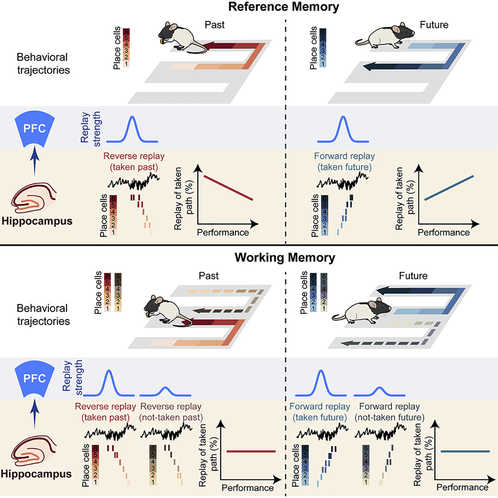
Dynamics of Awake Hippocampal-Prefrontal Replay for Spatial Learning and Memory-Guided Decision Making

Justin D.Shin, Wenbo Tang, Shantanu P. Jadhav

Spatial learning requires remembering and choosing paths to goals. Hippocampal place cells replay spatial paths during immobility in reverse and forward order, offering a potential mechanism. However, how replay supports both goal-directed learning and memory-guided decision making is unclear. We therefore continuously tracked awake replay in the same hippocampal-prefrontal ensembles throughout learning of a spatial alternation task. We found that, during pauses between behavioral trajectories, reverse and forward hippocampal replay supports an internal cognitive search of available past and future possibilities and exhibits opposing learning gradients for prediction of past and future behavioral paths, respectively. Coordinated hippocampal-prefrontal replay distinguished correct past and future paths from alternative choices, suggesting a role in recall of past paths to guide planning of future decisions for spatial working memory. Our findings reveal a learning shift from hippocampal reverse-replay-based retrospective evaluation to forward-replay-based prospective planning, with prefrontal readout of memory-guided paths for learning and decision making.

Convolutional Neural Networks as a Model of the Visual System: Past, Present, and Future

Grace W. Lindsay

Convolutional neural networks (CNNs) were inspired by early findings in the study of biological vision. They have since become successful tools in computer vision and state-of-the-art models of both neural activity and behavior on visual tasks. This review highlights what, in the context of CNNs, it means to be a good model in computational neuroscience and the various ways models can provide insight. Specifically, it covers the origins of CNNs and the methods by which we validate them as models of biological vision. It then goes on to elaborate on what we can learn about biological vision by understanding and experimenting on CNNs and discusses emerging opportunities for the use of CNNS in vision research beyond basic object recognition.